- Diet
- Cancer
- Colorectal Cancer
- Prostate Cancer
- Breast Cancer
- Adenoid Cystic Carcinoma
- Amyloidosis
- Anal Cancer
- Appendix Cancer
- Astrocytoma - Childhood
- Ataxia-Telangiectasia
- Beckwith-Wiedemann Syndrome
- Bile Duct Cancer (Cholangiocarcinoma)
- Birt-Hogg-Dubé Syndrome
- Bladder Cancer
- Bone Cancer (Sarcoma of Bone)
- Brain Stem Glioma - Childhood
- Brain Tumor
- Breast Cancer - Inflammatory
- Breast Cancer - Metastatic
- Breast Cancer - Male
- Carney Complex
- Central Nervous System Tumors (Brain and Spinal Cord) - Childhood
- Cervical Cancer
- Childhood Cancer
- Cowden Syndrome
- Craniopharyngioma - Childhood
- Desmoid Tumor
- Desmoplastic Infantile Ganglioglioma, Childhood Tumor
- Ependymoma - Childhood
- Esophageal Cancer
- Ewing Sarcoma - Childhood and Adolescence
- Eye Melanoma
- Eyelid Cancer
- Familial Adenomatous Polyposis
- Familial GIST
- Familial Malignant Melanoma
- Familial Pancreatic Cancer
- Gallbladder Cancer
- Gastrointestinal Stromal Tumor - GIST
- Germ Cell Tumor - Childhood
- Gestational Trophoblastic Disease
- Head and Neck Cancer
- Hereditary Breast and Ovarian Cancer
- Hereditary Diffuse Gastric Cancer
- Hereditary Leiomyomatosis and Renal Cell Cancer
- Hereditary Mixed Polyposis Syndrome
- Hereditary Pancreatitis
- Hereditary Papillary Renal Carcinoma
- HIV/AIDS-Related Cancer
- Juvenile Polyposis Syndrome
- Kidney Cancer
- Laryngeal and Hypopharyngeal Cancer
- Leukemia - Acute Lymphoblastic - ALL - Childhood
- Leukemia - Acute Lymphocytic - ALL
- Leukemia - Acute Myeloid - AML
- Leukemia - Acute Myeloid - AML - Childhood
- Leukemia - B-cell Prolymphocytic Leukemia and Hairy Cell Leukemia
- Leukemia - Chronic Lymphocytic - CLL
- Leukemia - Chronic Myeloid - CML
- Leukemia - Chronic T-Cell Lymphocytic
- Leukemia - Eosinophilic
- Li-Fraumeni Syndrome
- Liver Cancer
- Lung Cancer - Non-Small Cell
- Lung Cancer - Small Cell
- Lymphoma - Hodgkin
- Lymphoma - Hodgkin - Childhood
- Lynch Syndrome
- Lymphoma - Non-Hodgkin - Childhood
- Lymphoma - Non-Hodgkin
- Mastocytosis
- Medulloblastoma - Childhood
- Melanoma
- Meningioma
- Mesothelioma
- Multiple Endocrine Neoplasia Type 1
- Multiple Endocrine Neoplasia Type 2
- Multiple Myeloma
- MUTYH (or MYH)-Associated Polyposis
- Myelodysplastic Syndromes - MDS
- Nasal Cavity and Paranasal Sinus Cancer
- Nasopharyngeal Cancer
- Neuroblastoma - Childhood
- Neuroendocrine Tumor of the Gastrointestinal Tract
- Neuroendocrine Tumor of the Lung
- Neuroendocrine Tumor of the Pancreas
- Neuroendocrine Tumors
- Neurofibromatosis Type 1
- Neurofibromatosis Type 2
- Nevoid Basal Cell Carcinoma Syndrome
- Oral and Oropharyngeal Cancer
- Osteosarcoma - Childhood and Adolescence
- Ovarian, Fallopian Tube, and Peritoneal Cancer
- Pancreatic Cancer
- Parathyroid Cancer
- Penile Cancer
- Peutz-Jeghers Syndrome
- Pheochromocytoma and Paraganglioma
- Pituitary Gland Tumor
- Pleuropulmonary Blastoma - Childhood
- Retinoblastoma - Childhood
- Rhabdomyosarcoma - Childhood
- Salivary Gland Cancer
- Sarcoma - Kaposi
- Sarcomas, Soft Tissue
- Skin Cancer (Non-Melanoma)
- Small Bowel Cancer
- Stomach Cancer
- Testicular Cancer
- Thymoma and Thymic Carcinoma
- Thyroid Cancer
- Tuberous Sclerosis Complex
- Unknown Primary
- Uterine Cancer
- Vaginal Cancer
- Von Hippel-Lindau Syndrome
- Vulvar Cancer
- Waldenstrom Macroglobulinemia (Lymphoplasmacytic Lymphoma)
- Werner Syndrome
- Wilms Tumor - Childhood
- Xeroderma Pigmentosum
- Veterans with Cancer
- Insurance and Cancer
- Prayers for Cancer Healing
- Prayers for Cancer Survival
- Pharmacology - Cancer Oncology drugs
- Natural Cures for Cancer
- Cancer Causing Foods
- Cancer Fighting Foods
- Kaposi Sarcoma
- Nausea and Vomiting in Cancer
- Adrenocortical Carcinoma
- Adolescents and Young Adults with Cancer
- Basal Cell Carcinoma of the Skin
- Burkitt Lymphoma
- Pancreatic Cancer
- Pain Management in Cancer
- CBD and Cancer Patients
- Cancer Treatment
- Stoma Bag
- Cancer Bra
- Cancer Wigs
- Lymphedema and Cancer
- Ductal Carcinoma In Situ (DCIS)
- Mouth Cancer
- Pregnancy and Breast Cancer
- Endometrial Cancer
- Heart Tumors, Childhood
- Merkel Cell Carcinoma
- Urethral Cancer
- Cancer in Young Adults
- Exercise and Cancer
- Insurance Denial and Cancer
- Bronchial Tumors
- Colostomy and Cancer
- Tube Feeding and Cancer
- Chronic Myeloproliferative Neoplasms
- Pulmonary Inflammatory Myofibroblastic Tumor
- Cutaneous T-Cell Lymphoma
- Fallopian Tube Cancer
- Breast Prostheses after Mastectomy
- Vascular Tumors
- Urethral cancer
- Music
Sarcomas, Soft Tissue


In this video, Dr. Chetan Anchan (Orthopedic oncologist working at Speciality Surgical Oncology Hospital Mumbai) will explain to us how the diagnosis of Ewing’s Sarcoma is done.
Ewing Sarcoma, also known as PNET (Primitive Neuro Ectodermal Tumor), is a rare type of musculoskeletal cancer that can be found in bones as well as the muscle tissues around the bones. It is more commonly found in long bones such as the forearm bones or thigh bones. It is also found in pelvic bones, the spine, and ribs. It is found in neck muscles, thigh muscles, stomach muscles, and back muscles as well.
00:33 - How is Ewing Sarcoma diagnosed?
Ewing Sarcoma, like any other bone cancer, can be easily localized by doing an X-ray. It can be easily located and understood by the means of an X-ray. So when symptoms are found in the typical findings, there might be a chance of the presence of this disease.
After these findings are discovered and if these findings are paired with fever, pain, and swelling, doctors often get confused about it being osteomyelitis i.e. bone infection. Ewing’s Sarcoma is often confused to be osteomyelitis and is treated in the same way as osteomyelitis which can mar the treatment of Ewing’s Sarcoma. Thus only an X-ray and clinical symptoms are not enough for the diagnosis of Ewing’s Sarcoma.
An MRI scan is done and the entire organ that is affected by it is screened. MRIs can help to determine the exact extent of the tumor including nearby blood vessels and nerves. After confirming the presence of Ewing’s Sarcoma via X-ray and MRIs, a biopsy is done in which a sample is taken from the disease and sent to the lab to do further investigations and get a proper histopathological report.
To know more, watch the full video.
For any questions type them in the comment section.
__________________________
About Dr. Chetan Anchan
Dr. Chetan Anchan is an Orthopaedic surgeon from Mumbai. He has been practising Musculoskeletal Tumor Surgeon in Mumbai. He has experience in more than a thousand surgeries for bone and soft tissue tumors. With more than 16 years of specialty training/experience and practice, he provides the best patients care and provides them with the best advice and treatment.
Follow us on
Facebook : https://www.facebook.com/Dr-Ch....etan-Anchan-10588807
Instagram : https://instagram.com/dr_cheta....nanchan?utm_medium=c
Website : https://www.bonetumorclinic.in/
Thanks !!
#EwingSarcoma #cancerinchildren #diagnosi #drchetananchan #orthopediconcologist


For more information visit https://www.bonecancer.in/2020..../05/01/giant-cell-tu
To book an appointment with our team of experts, call (091) 9113940735
Find our complete library of success stories in orthopedic oncology https://youtube.com/playlist?l....ist=PLl20586fBuf6P0l
Our mission is to spread awareness on these rare diseases.
This is a short treatment outcome awareness video of a limb salvage surgery for soft tissue cancer by Dr Srimanth B S.
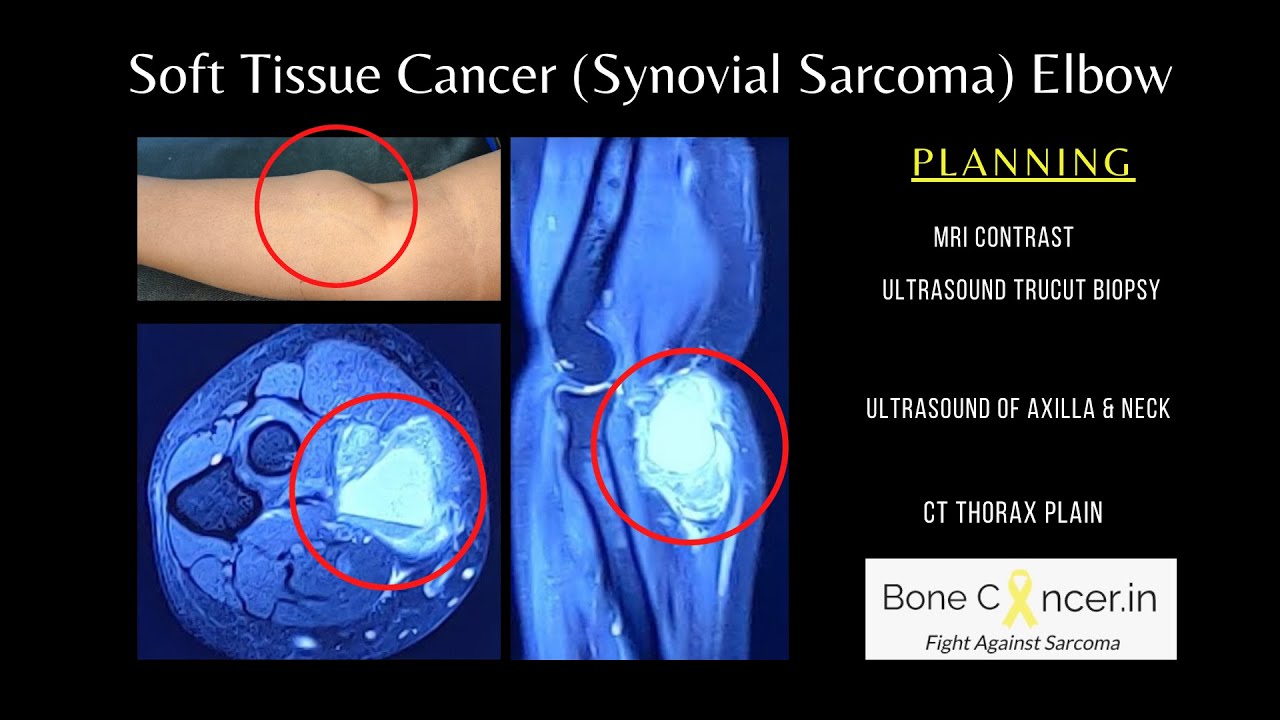
Synovial sarcoma is an aggressive soft tissue cancer affecting the adults. Ity can be seen commonly in limbs.
An image guided biopsy is performed to obtain the diagnosis. This is followed by Pet CT scan to stage and rule out distant metastases.
A multi disciplinary approach (Tumour Board) proposed personalized treatment plan. The lady underwent Tumor Excision and followed up with Adjuvant Radiotherapy to operated site. All vital nerves and blood vessels in elbow were saved to retain her hand.
Follow for more updates:
https://www.linkedin.com/in/srimanth-b-s-3634a766
https://instagram.com/musculos....keletal_oncology?igs
https://www.facebook.com/bonecancer.in/
#sarcoma #softtissuecancer #synovialsarcoma #limbsalvage #cancer #bonecancer #bonesarcoma #orthpaediconcology #symptoms #signs #treatment #medicaltourism


