- Diet
- Cancer
- Colorectal Cancer
- Prostate Cancer
- Breast Cancer
- Adenoid Cystic Carcinoma
- Amyloidosis
- Anal Cancer
- Appendix Cancer
- Astrocytoma - Childhood
- Ataxia-Telangiectasia
- Beckwith-Wiedemann Syndrome
- Bile Duct Cancer (Cholangiocarcinoma)
- Birt-Hogg-Dubé Syndrome
- Bladder Cancer
- Bone Cancer (Sarcoma of Bone)
- Brain Stem Glioma - Childhood
- Brain Tumor
- Breast Cancer - Inflammatory
- Breast Cancer - Metastatic
- Breast Cancer - Male
- Carney Complex
- Central Nervous System Tumors (Brain and Spinal Cord) - Childhood
- Cervical Cancer
- Childhood Cancer
- Cowden Syndrome
- Craniopharyngioma - Childhood
- Desmoid Tumor
- Desmoplastic Infantile Ganglioglioma, Childhood Tumor
- Ependymoma - Childhood
- Esophageal Cancer
- Ewing Sarcoma - Childhood and Adolescence
- Eye Melanoma
- Eyelid Cancer
- Familial Adenomatous Polyposis
- Familial GIST
- Familial Malignant Melanoma
- Familial Pancreatic Cancer
- Gallbladder Cancer
- Gastrointestinal Stromal Tumor - GIST
- Germ Cell Tumor - Childhood
- Gestational Trophoblastic Disease
- Head and Neck Cancer
- Hereditary Breast and Ovarian Cancer
- Hereditary Diffuse Gastric Cancer
- Hereditary Leiomyomatosis and Renal Cell Cancer
- Hereditary Mixed Polyposis Syndrome
- Hereditary Pancreatitis
- Hereditary Papillary Renal Carcinoma
- HIV/AIDS-Related Cancer
- Juvenile Polyposis Syndrome
- Kidney Cancer
- Laryngeal and Hypopharyngeal Cancer
- Leukemia - Acute Lymphoblastic - ALL - Childhood
- Leukemia - Acute Lymphocytic - ALL
- Leukemia - Acute Myeloid - AML
- Leukemia - Acute Myeloid - AML - Childhood
- Leukemia - B-cell Prolymphocytic Leukemia and Hairy Cell Leukemia
- Leukemia - Chronic Lymphocytic - CLL
- Leukemia - Chronic Myeloid - CML
- Leukemia - Chronic T-Cell Lymphocytic
- Leukemia - Eosinophilic
- Li-Fraumeni Syndrome
- Liver Cancer
- Lung Cancer - Non-Small Cell
- Lung Cancer - Small Cell
- Lymphoma - Hodgkin
- Lymphoma - Hodgkin - Childhood
- Lynch Syndrome
- Lymphoma - Non-Hodgkin - Childhood
- Lymphoma - Non-Hodgkin
- Mastocytosis
- Medulloblastoma - Childhood
- Melanoma
- Meningioma
- Mesothelioma
- Multiple Endocrine Neoplasia Type 1
- Multiple Endocrine Neoplasia Type 2
- Multiple Myeloma
- MUTYH (or MYH)-Associated Polyposis
- Myelodysplastic Syndromes - MDS
- Nasal Cavity and Paranasal Sinus Cancer
- Nasopharyngeal Cancer
- Neuroblastoma - Childhood
- Neuroendocrine Tumor of the Gastrointestinal Tract
- Neuroendocrine Tumor of the Lung
- Neuroendocrine Tumor of the Pancreas
- Neuroendocrine Tumors
- Neurofibromatosis Type 1
- Neurofibromatosis Type 2
- Nevoid Basal Cell Carcinoma Syndrome
- Oral and Oropharyngeal Cancer
- Osteosarcoma - Childhood and Adolescence
- Ovarian, Fallopian Tube, and Peritoneal Cancer
- Pancreatic Cancer
- Parathyroid Cancer
- Penile Cancer
- Peutz-Jeghers Syndrome
- Pheochromocytoma and Paraganglioma
- Pituitary Gland Tumor
- Pleuropulmonary Blastoma - Childhood
- Retinoblastoma - Childhood
- Rhabdomyosarcoma - Childhood
- Salivary Gland Cancer
- Sarcoma - Kaposi
- Sarcomas, Soft Tissue
- Skin Cancer (Non-Melanoma)
- Small Bowel Cancer
- Stomach Cancer
- Testicular Cancer
- Thymoma and Thymic Carcinoma
- Thyroid Cancer
- Tuberous Sclerosis Complex
- Unknown Primary
- Uterine Cancer
- Vaginal Cancer
- Von Hippel-Lindau Syndrome
- Vulvar Cancer
- Waldenstrom Macroglobulinemia (Lymphoplasmacytic Lymphoma)
- Werner Syndrome
- Wilms Tumor - Childhood
- Xeroderma Pigmentosum
- Veterans with Cancer
- Insurance and Cancer
- Prayers for Cancer Healing
- Prayers for Cancer Survival
- Pharmacology - Cancer Oncology drugs
- Natural Cures for Cancer
- Cancer Causing Foods
- Cancer Fighting Foods
- Kaposi Sarcoma
- Nausea and Vomiting in Cancer
- Adrenocortical Carcinoma
- Adolescents and Young Adults with Cancer
- Basal Cell Carcinoma of the Skin
- Burkitt Lymphoma
- Pancreatic Cancer
- Pain Management in Cancer
- CBD and Cancer Patients
- Cancer Treatment
- Stoma Bag
- Cancer Bra
- Cancer Wigs
- Lymphedema and Cancer
- Ductal Carcinoma In Situ (DCIS)
- Mouth Cancer
- Pregnancy and Breast Cancer
- Endometrial Cancer
- Heart Tumors, Childhood
- Merkel Cell Carcinoma
- Urethral Cancer
- Cancer in Young Adults
- Exercise and Cancer
- Insurance Denial and Cancer
- Bronchial Tumors
- Colostomy and Cancer
- Tube Feeding and Cancer
- Chronic Myeloproliferative Neoplasms
- Pulmonary Inflammatory Myofibroblastic Tumor
- Cutaneous T-Cell Lymphoma
- Fallopian Tube Cancer
- Breast Prostheses after Mastectomy
- Vascular Tumors
- Urethral cancer
- Music
Salivary Gland Cancer


For just $1/month, you can help keep these videos free! Subscribe to my Patreon at http://www.patreon.com/pwbmd
(Disclaimer: The medical information contained herein is intended for physician medical licensing exam review purposes only, and are not intended for diagnosis of any illness. If you think you may be suffering from any medical condition, you should consult your physician or seek immediate medical attention.)

http://www.EntHeadNeck.com.
http://www.nosesinus.com/
Dr Kevin Soh describes how to minimise injury to the facial nerve during parotid gland surgery (parotidectomy) using a nerve integrity monitor or nerve stimulator.
3 Mount Elizabeth, #07-02, Mount Elizabeth Medical Centre, Singapore 228510
https://www.google.com.sg/maps..../place/Dr+Kevin+Soh,
If you have any comments, PLEASE do not be afraid to ask. Please SUBSCRIBE, SHARE, and COMMENT on this video.
If you prefer to read, rather than watch the video, here’s the transcript.
0:16 – Case Presentation: A 39 year old man presents with a right neck swelling for one year. It is 3 cm in size. Fine needle aspiration cytology (FNAC) was performed. Cytology showed adenocarcinoma. Fine needle cytology has an accuracy rate of 90%.
0:34 – CT scan of the Parotid Gland. The normal parotid gland is radiolucent on CT scan. The tumor lies in the posterior part of the superficial lobe of the right parotid gland.
0:49 – MRI scans: A 3 cm right parotid mass is detected. Notice that the tumor enhances intensely with gadolinium administration. This suggests that the tumor is malignant.
1:14 – Positron Emission Tomography scan (PET scan): Used to detect spread of cancer to other parts of the body. The parotid tumor had high metabolic activity levels, which suggests that it is a cancer. Fortunately for this patient, the PET scan did not detect any other areas of increased metabolic activity. PET scans do not provide good anatomical information. PET scans are combined with CT scans to provide anatomical correlation. PET/CT scan showed an area of high metabolic activity in the right parotid gland. But the rest of the body is clear of tumor spread.
1:50 – Next, we have to remove the tumor. The facial nerve (or seventh cranial nerve) supplies the muscles of facial expression. This includes the orbicularis oculi muscle which allows us to close our eyes. It also includes the orbicularis oris muscle with controls our lip movements.
2:19 – Anatomy of the Facial Nerve: The facial nerve is only one of twelve cranial nerves. The facial nerve leaves the brain and enters the temporal bone. It travels horizontally through the middle ear. It descends vertically through the mastoid bone. Then, it exits through the stylomastoid foramen, just next to the styloid process.
3:02 - At the undersurface (ventral aspect) of the right temporal bone, the styloid process is identified. The facial nerve exits the temporal bone at the stylomastoid foramen, behind the styloid process. After exiting the stylomastoid foramen, the facial nerve enters the parotid gland. The facial nerve divides the parotid gland into a small deep lobe and a large superficial lobe.
3:52 – The main trunk of the facial nerve first divides into 2 large branches: the zygomatico-temporal branch and the cervico-facial branch. The zygomatico-temporal branch gives rise to the temporal branch, zygomatic branch, and the buccal branches. The cervico-facial branch gives rise to the marginal mandibular branch, and the cervical branches.
4:08 – The facial nerve branches look like the feet of a goose. They are sometimes called PES ANSERINUS (or goose feet).
4:19 – NIM Response 2.0 Nerve Integrity Monitor. The nerve integrity monitor facilitates safe facial nerve identification and dissection.
4:37 – Demonstration of Parotidectomy Procedure: The incision is marked out carefully to hide the scar. The incision is planned along natural skin creases. NIM electrode placement into the orbicularis oculi muscle. This electrode monitors the temporal and zygomatic branches of the facial nerve. NIM electrode placement within the nasolabial groove into the orbicularis oris muscle. This monitors the buccal and marginal mandibular branches of the facial nerve. The nerve integrity monitor is then turned on. The electrodes are tested to ensure correct placement.
5:19 – The sub-platysmal skin flap is elevated. The skin flap is sutured down. The great auricular nerve is identified. We have to dissect here to look for the main trunk of facial nerve. We can now see the main trunk of the facial nerve.
6:58 – At the completion of parotid surgery, the nerve integrity monitor is used to confirm nerve integrity. Stimulate both proximally and distally to confirm nerve integrity. The stimulator checks the integrity of the main trunk of facial nerve. Then we check the upper branches and the lower branches.

This video describes treatment of Parotid and submandibular salivary gland infections. The video is for doctors and healthcare providers only. Please consult your doctor prior to following the recommendations in the video. For more information about Dr. Kakani, please visit www.licent.org


Salivary Gland Stones: Frequently Asked Questions. Head & Neck Surgical Oncologist, Dr. Ryan Osborne, Director of Head & Neck Surgery and Salivary Gland Disorders, at the Osborne Head & Neck Institute, specializes in the non-surgical removal of salivary gland stones causing swelling and infection of the major salivary glands. In this series of videos, Dr. Osborne answers frequently asked questions about salivary gland stones.
0:00:11 - What is a salivary gland stone?
0:00:26 - What causes salivary gland stones?
0:00:55 - What are the symptoms of a salivary stone?
0:01:17 - How is a salivary gland stone diagnosed?
0:01:37 - Do salivary gland stones need to be removed?
0:02:07 - Can a blocked salivary gland stone go away on its own?
0:02:27 - How do you get rid of salivary gland stones?
0:03:12 - Where does a salivary stone come out?
0:03:36 - How long do salivary gland stones last?
0:04:01 - How common are salivary gland stones?
0:04:32 - What is a blocked salivary gland?
0:04:55 - How long does it take to recover from salivary gland surgery?
0:05:34 - What are the symptoms of salivary gland cancer?
0:06:24 - What are the symptoms of a salivary gland tumor?
0:07:13 - Is salivary gland cancer curable?
For in depth information, or to schedule your virtual consultation with Dr. Osborne, please visit: https://www.parotid.net/
Watch surgical procedures, including salivary gland surgery, on our docu-series called OHNI Case Files: https://www.youtube.com/playli....st?list=PLD7nLk0Vkgv


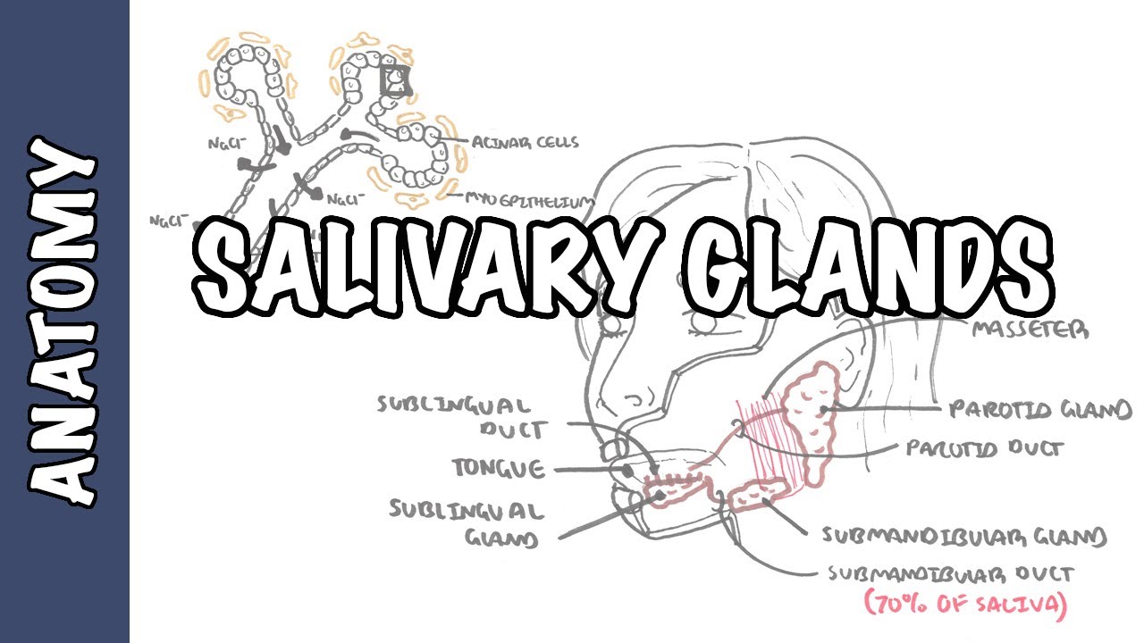
Buy Images here: armandoh.org/shop
"We have three pairs of salivary glands bilaterally. Despite the parotid gland being large, the submandibular gland produces majority of the saliva 70% second is the parotid gland 25%. Saliva is produced and secreted in the mouth producing an alkali substance. The functional unit of salivary glands is called a SALIVON."
Where do I get my information from: http://armandoh.org/resource
Facebook:
https://www.facebook.com/ArmandoHasudungan
Support me:
http://www.patreon.com/armando
Instagram:
http://instagram.com/armandohasudungan
Twitter:
https://twitter.com/Armando71021105
SPECIAL THANKS:
Patreon members
FaberCastell Australia - https://www.youtube.com/user/FaberCastellGroup
What markers do I use?
FaberCastellPITTartistpens1,5
FaberCastellPITTartistpensF
FaberCastellPermanentmarkers
FaberCastellPITTartistpensbrush

Eric Moore, M.D., a head and neck surgeon at Mayo Clinic, and Heidi, a Mayo Clinic patient, talk about what to expect with the testing ordered by your doctor.
Get Parotid Tumor Care Now. Call to obtain an appointment at Mayo Clinic, Otolaryngology-Head and Neck Surgery 1-507-923-2443. See here to learn more about Parotid Tumors https://mayocl.in/3cu5iCE
