- Diet
- Cancer
- Colorectal Cancer
- Prostate Cancer
- Breast Cancer
- Adenoid Cystic Carcinoma
- Amyloidosis
- Anal Cancer
- Appendix Cancer
- Astrocytoma - Childhood
- Ataxia-Telangiectasia
- Beckwith-Wiedemann Syndrome
- Bile Duct Cancer (Cholangiocarcinoma)
- Birt-Hogg-Dubé Syndrome
- Bladder Cancer
- Bone Cancer (Sarcoma of Bone)
- Brain Stem Glioma - Childhood
- Brain Tumor
- Breast Cancer - Inflammatory
- Breast Cancer - Metastatic
- Breast Cancer - Male
- Carney Complex
- Central Nervous System Tumors (Brain and Spinal Cord) - Childhood
- Cervical Cancer
- Childhood Cancer
- Cowden Syndrome
- Craniopharyngioma - Childhood
- Desmoid Tumor
- Desmoplastic Infantile Ganglioglioma, Childhood Tumor
- Ependymoma - Childhood
- Esophageal Cancer
- Ewing Sarcoma - Childhood and Adolescence
- Eye Melanoma
- Eyelid Cancer
- Familial Adenomatous Polyposis
- Familial GIST
- Familial Malignant Melanoma
- Familial Pancreatic Cancer
- Gallbladder Cancer
- Gastrointestinal Stromal Tumor - GIST
- Germ Cell Tumor - Childhood
- Gestational Trophoblastic Disease
- Head and Neck Cancer
- Hereditary Breast and Ovarian Cancer
- Hereditary Diffuse Gastric Cancer
- Hereditary Leiomyomatosis and Renal Cell Cancer
- Hereditary Mixed Polyposis Syndrome
- Hereditary Pancreatitis
- Hereditary Papillary Renal Carcinoma
- HIV/AIDS-Related Cancer
- Juvenile Polyposis Syndrome
- Kidney Cancer
- Laryngeal and Hypopharyngeal Cancer
- Leukemia - Acute Lymphoblastic - ALL - Childhood
- Leukemia - Acute Lymphocytic - ALL
- Leukemia - Acute Myeloid - AML
- Leukemia - Acute Myeloid - AML - Childhood
- Leukemia - B-cell Prolymphocytic Leukemia and Hairy Cell Leukemia
- Leukemia - Chronic Lymphocytic - CLL
- Leukemia - Chronic Myeloid - CML
- Leukemia - Chronic T-Cell Lymphocytic
- Leukemia - Eosinophilic
- Li-Fraumeni Syndrome
- Liver Cancer
- Lung Cancer - Non-Small Cell
- Lung Cancer - Small Cell
- Lymphoma - Hodgkin
- Lymphoma - Hodgkin - Childhood
- Lynch Syndrome
- Lymphoma - Non-Hodgkin - Childhood
- Lymphoma - Non-Hodgkin
- Mastocytosis
- Medulloblastoma - Childhood
- Melanoma
- Meningioma
- Mesothelioma
- Multiple Endocrine Neoplasia Type 1
- Multiple Endocrine Neoplasia Type 2
- Multiple Myeloma
- MUTYH (or MYH)-Associated Polyposis
- Myelodysplastic Syndromes - MDS
- Nasal Cavity and Paranasal Sinus Cancer
- Nasopharyngeal Cancer
- Neuroblastoma - Childhood
- Neuroendocrine Tumor of the Gastrointestinal Tract
- Neuroendocrine Tumor of the Lung
- Neuroendocrine Tumor of the Pancreas
- Neuroendocrine Tumors
- Neurofibromatosis Type 1
- Neurofibromatosis Type 2
- Nevoid Basal Cell Carcinoma Syndrome
- Oral and Oropharyngeal Cancer
- Osteosarcoma - Childhood and Adolescence
- Ovarian, Fallopian Tube, and Peritoneal Cancer
- Pancreatic Cancer
- Parathyroid Cancer
- Penile Cancer
- Peutz-Jeghers Syndrome
- Pheochromocytoma and Paraganglioma
- Pituitary Gland Tumor
- Pleuropulmonary Blastoma - Childhood
- Retinoblastoma - Childhood
- Rhabdomyosarcoma - Childhood
- Salivary Gland Cancer
- Sarcoma - Kaposi
- Sarcomas, Soft Tissue
- Skin Cancer (Non-Melanoma)
- Small Bowel Cancer
- Stomach Cancer
- Testicular Cancer
- Thymoma and Thymic Carcinoma
- Thyroid Cancer
- Tuberous Sclerosis Complex
- Unknown Primary
- Uterine Cancer
- Vaginal Cancer
- Von Hippel-Lindau Syndrome
- Vulvar Cancer
- Waldenstrom Macroglobulinemia (Lymphoplasmacytic Lymphoma)
- Werner Syndrome
- Wilms Tumor - Childhood
- Xeroderma Pigmentosum
- Veterans with Cancer
- Insurance and Cancer
- Prayers for Cancer Healing
- Prayers for Cancer Survival
- Pharmacology - Cancer Oncology drugs
- Natural Cures for Cancer
- Cancer Causing Foods
- Cancer Fighting Foods
- Kaposi Sarcoma
- Nausea and Vomiting in Cancer
- Adrenocortical Carcinoma
- Adolescents and Young Adults with Cancer
- Basal Cell Carcinoma of the Skin
- Burkitt Lymphoma
- Pancreatic Cancer
- Pain Management in Cancer
- CBD and Cancer Patients
- Cancer Treatment
- Stoma Bag
- Cancer Bra
- Cancer Wigs
- Lymphedema and Cancer
- Ductal Carcinoma In Situ (DCIS)
- Mouth Cancer
- Pregnancy and Breast Cancer
- Endometrial Cancer
- Heart Tumors, Childhood
- Merkel Cell Carcinoma
- Urethral Cancer
- Cancer in Young Adults
- Exercise and Cancer
- Insurance Denial and Cancer
- Bronchial Tumors
- Colostomy and Cancer
- Tube Feeding and Cancer
- Chronic Myeloproliferative Neoplasms
- Pulmonary Inflammatory Myofibroblastic Tumor
- Cutaneous T-Cell Lymphoma
- Fallopian Tube Cancer
- Breast Prostheses after Mastectomy
- Vascular Tumors
- Urethral cancer
- Music
Ependymoma - Childhood

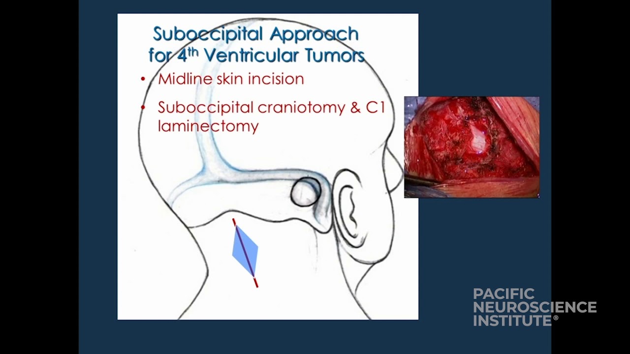

Ependymoma Surgery in the 4th ventricle requires microsurgical and endoscopic techniques for safe and maximal tumor resection.
In this video, Dr. Daniel Kelly, Director of the Pacific Brain Tumor Center, Providence Saint John's Health Center demonstrates the surgical removal of an ependymoma within the 4th ventricle using microsurgical techniques and endoscopic visualization.
Ependymomas are glial neoplasms (gliomas) that can arise in any of the brain's fluid-filled chambers (cerebral ventricles). When they arise in the 4th ventricle, they can cause headaches, slurred speech, swallowing difficulties, coordination and balance difficulties as well as hydrocephalus. In this case, the patient was a young man who developed progressive and severe headaches leading to an MRI and the diagnosis of a 4th ventricular tumor. The MRI clearly showed the tumor causing pressure on the brainstem and cerebellum. The surgical video demonstrates how the tumor was approached through a suboccipital craniotomy, the route into the 4th ventricle, microscopic tumor removal and finally the use of endoscopy to confirm maximal tumor resection. Fortunately in our patient, over 97% of the tumor was able to be removed and it was low grade ependymomas (WHO Grade II). Given the small remaining tumor remnant left adherent to the brainstem, he was treated with stereotactic radiation shortly after surgery. Now more than 5 years after surgery, he is doing well with no evidence of tumor regrowth.
At the Pacific Brain Tumor Center we have one of the world's largest experiences treating all types of brain and skull base tumors using minimally invasive keyhole and endoscopic approaches.
https://pacificneuro.org
https://pacificneuro.org/kelly
https//pacificbraintumor.org | 310-582-7450
We are IndianRadiologist - www.indianradiologist.com
Follow us on Social Media for Event info, New videos, Free Classifieds Information on Jobs & Machines, Unusual & Rare Radiology Images, New Product Reviews & More
YOUTUBE: Subscribe & Click on the Bell Icon for notifications: https://www.youtube.com/indian....radiologist?sub_conf
FACEBOOK: https://www.facebook.com/groups/indianradiologist/
YOUTUBE: www.youtube.com/indianradiologist
INSTAGRAM: https://www.instagram/Indianradiologist
#braintumor #mribraintumour #ependymoma
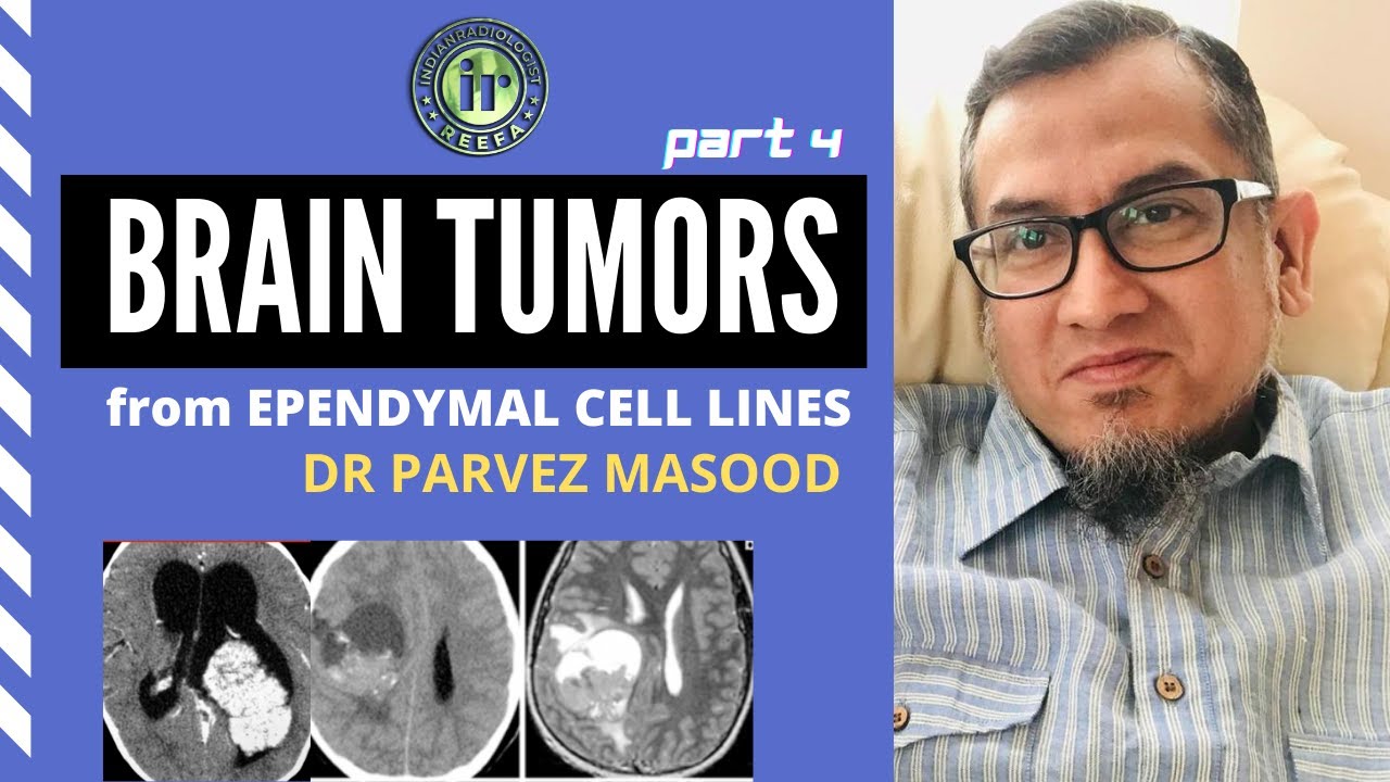
In this video, learn about: ependymoma, subependymoma, hydrocephalus, mass effect, choroid plexus tumors.
Quick learning videos on Radiology for UG and Residents in Radiology. Subscribe to Indian Radiologist and get free Radiology teaching videos from experts in the field of Radiology.

There are multiple treatment paths for patients with ependymoma. The CERN Foundation has created an international clinical trials network that includes several clinical centers of excellence in both pediatric and adult ependymomas in the United States. Currently, the CERN Foundation is developing clinical trials designed for ependymoma patients. Patients that meet eligibility requirements must join the trial at a CERN Center. We are pleased to have two open trials, one for adults with ependymoma and one for children with ependymoma, and expect to open additional trials in the future. Learn more about ependymoma and the CERN Foundation at www.cern-foundation.org. You can join our e-newsletter list to receive updates on ependymoma and join the message boards.

Aimee Garrison was 26 weeks pregnant when the pain became overwhelming. She had trouble sleeping and keeping up with her toddler. An MRI revealed a rare spinal cord ependymoma, which affects less than 2,000 adults each year. A tumor the size of a baby carrot had been slowly growing in Aimee’s spinal cord, pushing it against her vertebrae.
Learn more about Aimee: http://uofmhealthblogs.org/neu....rohealth/marathon-ru

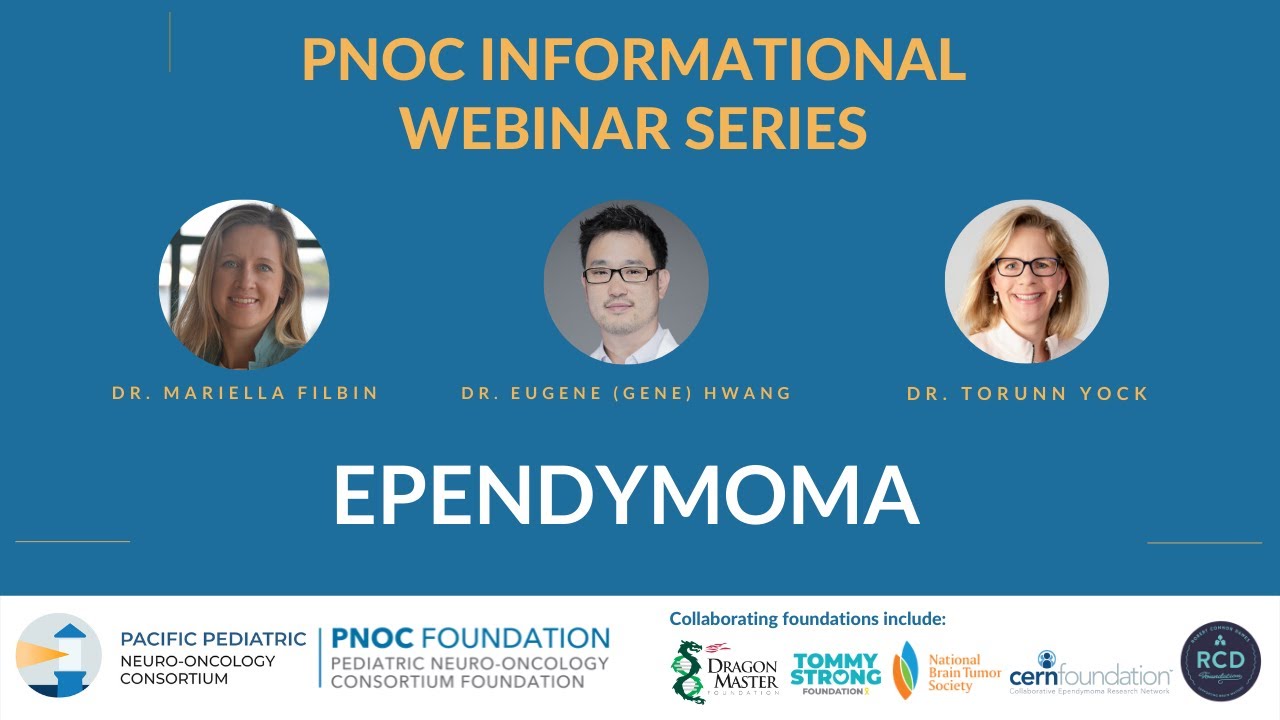
The Pacific Pediatric Neuro-Oncology Consortium (PNOC) is an international consortium, led by Dr. Sabine Mueller and Dr. Michael Prados, with centers within the United States, Europe, Asia and Australia. PNOC are dedicated to bringing new therapies to children and young adults with brain tumors. PNOC's goal is to improve outcomes by translating the latest findings in brain tumor biology into better treatments for these children.
As part of our ongoing webinar series on May 21st we presented the PNOC Ependymoma webinar to provide updates on research and clinical trials and to answer patient family questions.
FEATURED SPEAKERS:
Dr. Mariella Filbin Co-Director for Research, Pediatric Neuro-oncology Program, Assistant Professor of Pediatrics, Harvard Medical School; Associate Member, Broad Institute of Harvard and MIT, Dana-Farber/Boston Children's Cancer and Blood Disorder Center
Dr. Eugene (Gene) Hwang Pediatric Neuro-Oncologist, PNOC Ependymoma Research Group; Associate Division Chief, Oncology, Children's National Hospital
Dr. Torunn Yock Director, Pediatric Radiation Oncology, MGH; Professor, Harvard Medical School; Chair, Quality Improvement Committee, Francis H. Burr Proton Therapy Center
and
Dr. Sabine Mueller PNOC Co-Founder and Project Leader, Professor of Clinical Neurology, UCSF
Dr. Cassie Kline Attending Physician, Director, Neuro-Oncology Clinical Research; Kortney Rose Foundation Clinical Researcher in Neuro-Oncology; Children’s Hospital of Philadelphia; PNOC Director of Data Quality and Integration
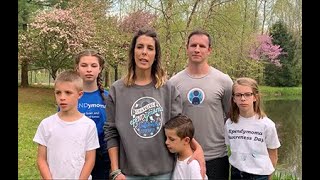
Thank you to Lindsay for sharing her story today and all the patient families who tuned in live and asked questions.
For more information on PNOC and PNOC's Ependymoma Research Group please visit https://pnoc.us
If you would like to learn more about PNOC Foundation or support Ependymoma research please visit https://www.pnocfoundation.org
PNOC is not only a collaboration of doctors, scientists and patient families, it also represents a collaboration of funders and advocates. We gratefully acknowledge the support of PNOC's Ependymoma work and this webinar by following collaborations foundations: Dragon Master Foundation, Tommy Strong Foundation, National Brain Tumor Society, Robert Dawes Foundation and CERN Foundation.