- Diet
- Cancer
- Colorectal Cancer
- Prostate Cancer
- Breast Cancer
- Adenoid Cystic Carcinoma
- Amyloidosis
- Anal Cancer
- Appendix Cancer
- Astrocytoma - Childhood
- Ataxia-Telangiectasia
- Beckwith-Wiedemann Syndrome
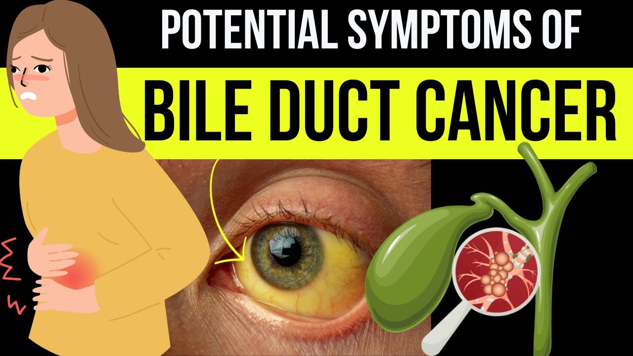
- Bile Duct Cancer (Cholangiocarcinoma)
- Birt-Hogg-Dubé Syndrome
- Bladder Cancer
- Bone Cancer (Sarcoma of Bone)
- Brain Stem Glioma - Childhood
- Brain Tumor
- Breast Cancer - Inflammatory
- Breast Cancer - Metastatic
- Breast Cancer - Male
- Carney Complex
- Central Nervous System Tumors (Brain and Spinal Cord) - Childhood
- Cervical Cancer
- Childhood Cancer
- Cowden Syndrome
- Craniopharyngioma - Childhood
- Desmoid Tumor
- Desmoplastic Infantile Ganglioglioma, Childhood Tumor
- Ependymoma - Childhood
- Esophageal Cancer
- Ewing Sarcoma - Childhood and Adolescence
- Eye Melanoma
- Eyelid Cancer
- Familial Adenomatous Polyposis
- Familial GIST
- Familial Malignant Melanoma
- Familial Pancreatic Cancer
- Gallbladder Cancer
- Gastrointestinal Stromal Tumor - GIST
- Germ Cell Tumor - Childhood
- Gestational Trophoblastic Disease
- Head and Neck Cancer
- Hereditary Breast and Ovarian Cancer
- Hereditary Diffuse Gastric Cancer
- Hereditary Leiomyomatosis and Renal Cell Cancer
- Hereditary Mixed Polyposis Syndrome
- Hereditary Pancreatitis
- Hereditary Papillary Renal Carcinoma
- HIV/AIDS-Related Cancer
- Juvenile Polyposis Syndrome
- Kidney Cancer
- Laryngeal and Hypopharyngeal Cancer
- Leukemia - Acute Lymphoblastic - ALL - Childhood
- Leukemia - Acute Lymphocytic - ALL
- Leukemia - Acute Myeloid - AML
- Leukemia - Acute Myeloid - AML - Childhood
- Leukemia - B-cell Prolymphocytic Leukemia and Hairy Cell Leukemia
- Leukemia - Chronic Lymphocytic - CLL
- Leukemia - Chronic Myeloid - CML
- Leukemia - Chronic T-Cell Lymphocytic
- Leukemia - Eosinophilic
- Li-Fraumeni Syndrome
- Liver Cancer
- Lung Cancer - Non-Small Cell
- Lung Cancer - Small Cell
- Lymphoma - Hodgkin
- Lymphoma - Hodgkin - Childhood
- Lynch Syndrome
- Lymphoma - Non-Hodgkin - Childhood
- Lymphoma - Non-Hodgkin
- Mastocytosis
- Medulloblastoma - Childhood
- Melanoma
- Meningioma
- Mesothelioma
- Multiple Endocrine Neoplasia Type 1
- Multiple Endocrine Neoplasia Type 2
- Multiple Myeloma
- MUTYH (or MYH)-Associated Polyposis
- Myelodysplastic Syndromes - MDS
- Nasal Cavity and Paranasal Sinus Cancer
- Nasopharyngeal Cancer
- Neuroblastoma - Childhood
- Neuroendocrine Tumor of the Gastrointestinal Tract
- Neuroendocrine Tumor of the Lung
- Neuroendocrine Tumor of the Pancreas
- Neuroendocrine Tumors
- Neurofibromatosis Type 1
- Neurofibromatosis Type 2
- Nevoid Basal Cell Carcinoma Syndrome
- Oral and Oropharyngeal Cancer
- Osteosarcoma - Childhood and Adolescence
- Ovarian, Fallopian Tube, and Peritoneal Cancer
- Pancreatic Cancer
- Parathyroid Cancer
- Penile Cancer
- Peutz-Jeghers Syndrome
- Pheochromocytoma and Paraganglioma
- Pituitary Gland Tumor
- Pleuropulmonary Blastoma - Childhood
- Retinoblastoma - Childhood
- Rhabdomyosarcoma - Childhood
- Salivary Gland Cancer
- Sarcoma - Kaposi
- Sarcomas, Soft Tissue
- Skin Cancer (Non-Melanoma)
- Small Bowel Cancer
- Stomach Cancer
- Testicular Cancer
- Thymoma and Thymic Carcinoma
- Thyroid Cancer
- Tuberous Sclerosis Complex
- Unknown Primary
- Uterine Cancer
- Vaginal Cancer
- Von Hippel-Lindau Syndrome
- Vulvar Cancer
- Waldenstrom Macroglobulinemia (Lymphoplasmacytic Lymphoma)
- Werner Syndrome
- Wilms Tumor - Childhood
- Xeroderma Pigmentosum
- Veterans with Cancer
- Insurance and Cancer
- Prayers for Cancer Healing
- Prayers for Cancer Survival
- Pharmacology - Cancer Oncology drugs
- Natural Cures for Cancer
- Cancer Causing Foods
- Cancer Fighting Foods
- Kaposi Sarcoma
- Nausea and Vomiting in Cancer
- Adrenocortical Carcinoma
- Adolescents and Young Adults with Cancer
- Basal Cell Carcinoma of the Skin
- Burkitt Lymphoma
- Pancreatic Cancer
- Pain Management in Cancer
- CBD and Cancer Patients
- Cancer Treatment
- Stoma Bag
- Cancer Bra
- Cancer Wigs
- Lymphedema and Cancer
- Ductal Carcinoma In Situ (DCIS)
- Mouth Cancer
- Pregnancy and Breast Cancer
- Endometrial Cancer
- Heart Tumors, Childhood
- Merkel Cell Carcinoma
- Urethral Cancer
- Cancer in Young Adults
- Exercise and Cancer
- Insurance Denial and Cancer
- Bronchial Tumors
- Colostomy and Cancer
- Tube Feeding and Cancer
- Chronic Myeloproliferative Neoplasms
- Pulmonary Inflammatory Myofibroblastic Tumor
- Cutaneous T-Cell Lymphoma
- Fallopian Tube Cancer
- Breast Prostheses after Mastectomy
- Vascular Tumors
- Urethral cancer
- Music


![Imaging of bile duct inflammation - HD [Basic Radiology]](https://i.ytimg.com/vi/P5QmaGr-0NY/mqdefault.jpg)


Bile Duct Ultrasound Normal Vs Abnormal Image Appearances | Biliary Tract Abnormalities USG Scan

Bile Duct Ultrasound Normal Vs Abnormal Image Appearances | Biliary Tract Abnormalities USG Scan
Intro - 0:00
Normal Bile Duct - 0:06
Fusiform Dilation (Choledochal Cyst) - 1:35
Dilated Common Bile Duct - 2:03
Caroli Disease - 2:28
Central Dot Sign - 3:09
Intrahepatic Biliary Stones - 3:29
Common Bile Duct Stones - 3:43
Mirizzi Syndrome - 3:51
Hemobilia - 4:18
Pneumobilia - 5:02
Acute Cholangitis - 5:23
Recurrent Pyogenic Cholangitis - 6:08
Primary Sclerosing Cholangitis - 6:31
Ascariasis - 6:50
HIV (AIDS) Cholangiopathy - 7:16
Intraductal Cholangiocarcinoma - 7:36
Intrahepatic Cholangiocarcinoma - 7:59
Hilar Cholangiocarcinoma - 8:18
You can support our work with a donation:
Airtm: drsamslibrary@gmail.com
Patreon: patreon.com/drsamsimaginglibrary
Guidelines On How To Read And Understand My Videos:
1. Click on the video of your choice.
2. The normal organ images will appear on the left half of the screen.
3. The images with pathologies/diseases will be displayed on the right half of the image.
4. Each case image has description explaining the features of the pathology.
5. Please turn on Subtitles/Closed Caption for easier understanding of the audio, or if you are watching the video on mute.
6. The subtitles may overlap and cover the description. You can move the subtitles away from the description by dragging it with the cursor or with finger touch on smartphone.
Related: images, cases, scans, imaging, pathologies, abnormalities, radiology, sonography, ultrasonography, reporting, categorization, snowstorm, hyperechoic, isoechoic, hypoechoic, calcifications, diseases, cancer, stones, tree, cbd, common hepatic duct, diameter

SORT BY-
Top Comments
-
Latest comments