Laatste video's


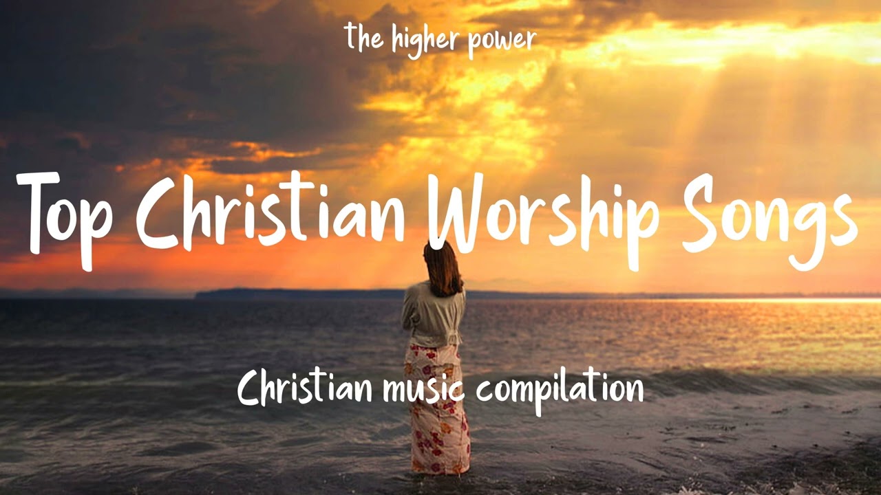

Top Christian Worship Songs 2023 ~ Playlist Hillsong Praise & Worship Songs
#christianworshipsongs #hillsongworship #TheHigherPower #Christian #ChristianMusic
Our socials network:
The Higher Power playlist: https://artlnk.info/TheHigherPower_playlist
Spotify: https://sptlnk.com/TheHigherPower
Facebook: http://bit.ly/fbhigherpower
"Let everything that has breath praise the Lord. Praise the Lord." Psalm 150:6
God bless us!
#gospel #gospelsong #godmusic #gospelmusic #christiansonglyrics #praiseandworship #worshipsong #newmusic #praisesongs #praisingsongs #christiansong #thehigherpower #higherpower #churchjesus #gospellyrics #gospelsonglyrics #worshipsongs #lyrics #lyrical #Worshiplyrics #Jesus #Jesussongs #songofworship #God #christian #2022 #Christ #jesus #christ #lyrics2022 #christiansongs #christiansongs2022 #christianmusic #worship #worshipsongs #hillsongworship #worshipsongs2022 #Christianmusicmix #Christiansongsplaylist
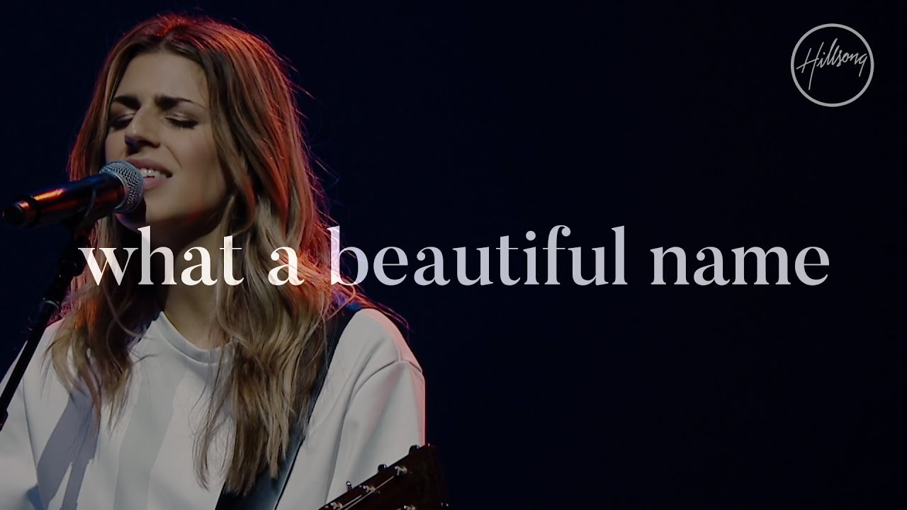
"What A Beautiful Name" from the album 'let there be light'' recorded live at Hillsong Conference in Sydney, 2016.
** Scriptural Inspiration Behind the Lyrics**
https://goo.gl/N5M5Qh
** Song Story From Brooke Ligertwood**
https://goo.gl/OqOJxU
**Subscribe to our YouTube channel**
https://worship.lnk.to/subscribe
Click to listen to Hillsong Worship's latest release, 'Team Night' here: https://worship.lnk.to/teamnightID
Click here to listen to the latest from Hillsong Worship: https://hillsong.lnk.to/ytplaylist
Experience the beauty of the gathering of The CHURCH from the comfort of your home.
Join in with an exclusive behind-the-scenes peak into Hillsong Conference from over the years at https://hillsongchannelnow.com/
Stay connected:
Instagram: https://instagram.com/hillsongworship
Facebook: https://facebook.com/hillsongworship
Twitter: https://twitter.com/hillsongworship
Website: https://hillsong.com/worship
What A Beautiful Name
Words and Music by Ben Fielding & Brooke Ligertwood
© 2016 Hillsong Music Publishing.
CCLI: 7068424
VERSE 1:
You were the Word at the beginning
One with God the Lord Most High
Your hidden glory in creation
Now revealed in You our Christ
CHORUS 1:
What a beautiful Name it is
What a beautiful Name it is
The Name of Jesus Christ my King
What a beautiful Name it is
Nothing compares to this
What a beautiful Name it is
The Name of Jesus
VERSE 2:
You didn’t want heaven without us
So Jesus You brought heaven down
My sin was great Your love was greater
What could separate us now
CHORUS 2:
What a wonderful Name it is
What a wonderful Name it is
The Name of Jesus Christ my King
What a wonderful Name it is
Nothing compares to this
What a wonderful Name it is
The Name of Jesus
What a wonderful Name it is
The Name of Jesus
BRIDGE:
Death could not hold You
The veil tore before You
You silence the boast of sin and grave
The heavens are roaring
The praise of Your glory
For You are raised to life again
You have no rival
You have no equal
Now and forever God You reign
Yours is the kingdom
Yours is the glory
Yours is the Name above all names
CHORUS 3:
What a powerful Name it is
What a powerful Name it is
The Name of Jesus Christ my King
What a powerful Name it is
Nothing can stand against
What a powerful Name it is
The Name of Jesus
TAGS:
What a powerful Name it is
The Name of Jesus
What a powerful Name it is
The Name of Jesus
#whatabeautifulname #hillsongworship #lettherebelight

Nonstop Praise And Worship Songs | Best 100 Praise And Worship Songs | Best Christian Songs 2023
---------------------------------------------------
🔻Follow "Praise Worship Music"
➞ Top Vidoes Hillsongs: https://bit.ly/3xbFxir
➞ Subscribe for More: https://bitly.com.vn/j6ar4y
✚Please share this video in social sites (Facebook, Google +, Twitter.)✚
📌 Facebook: https://bitly.com.vn/zvzxcl
📌 Soundcloud: https://bitly.com.vn/qgmtfj
📌 TikTok: https://bitly.com.vn/pthcga
📌 Instagram: https://bitly.com.vn/g75awh
We hope to hear your comments
#worshipsongs #christiangospel #praiseandworship
---------------------------------------------------
► Music and Video Copyright belongs to @Praise Worship Music. DO NOT reupload, otherwise, you will get copyright strikes.
Sincere thanks: Praise Worship Music Team !
---------------------------------------------------
►Tag:
worship,praise,praise and worship,,gospel music,gospel topic,worship songs,praise worship songs,worship and praise songs,worship songs 2021,gospel songs ,christian songs and praise,praise songs,christian songs 2022 ,latest gospel music,christian songs with lyrics,christian music,best worship songs gospel songs,christian songs ,worship songs with lyrics,praise and worship songs with lyrics
#praiseworshipsongs #praiseandworship #worship
#worshipsongs #BestPraiseWorship #praiseandworshipsongswithlyrics
#Praise
#Christian
#Gospel
#worshipsongswithlyrics
#worshipsongsofhillsong
#worshipsongslyrics
#worshipsongsever
#worshipsongscollection
#worshipsongsalltime
#worshipsongs
#worshipmusicchristian
#worshipmusic2021
#worshipmusic
#worshipgospelmusic
#worshipandpraisesongs
#worship2021
#worship
#wepraiseyou
#topworshipsongs
#toppraiseandworshipsongs
#top100praiseandworshipsongs2021
#top100christianworshipsongs
#top100bestchristiangospel
#prayers
#prayersongslyrics
#prayersongs2021
#prayersongs
#praiseworshipsongs
#praiseworshipmusic
#praisethelord
#Lordineedyou
#praisesongs
#praiselord
#praiseandworshipsongs2021
#praiseandworshipsongs
#praiseandworshipmusic
#praiseandworship
#praiseandhymns
#praise&worshipsongs2021
#praise
#powerfulpraiseandworshipsongs
#nonstopworshipsongs
#nonstopchristiansongs
#nigeriangospelmusic
#nigeriangospelmix
#newworshipsongs
#newpraiseandworshipsongs
#newchristiansongs
#newchristian
#musicpraise
#morningworshipsongs2021
#morningworshipsongs
#morningworship
#morningpraiseandworshipsongs
#morningworshipsongs
#louvoresdeadoração
#laurendaigleworshipsongs
#laurendaiglesongs
#laurendaigleplaylist
#laurendaiglefullalbum
#laurendaiglealbums
#laurendaigle
#latestpraisesongs
#latestgospelsongs2021
#latestgospelsongs
#latestgospelmusic
#latestchristiansongs2021
#latestchristiangospel
#hymnssongspraise
#hymnssongs
#hymnsoffaith
#hymns
#hillsongspraiseandworshipsongsplaylist
#hillsongworshiptoptracks
#hillsongworshipsongs
#hillsongworshiplyrics
#hillsongworship2021
#hillsongworship
#hillsongunited
#hillsongtoptracks
#hillsongsongslyrics
#hillsongsongs
#hillsongpraiseandworshipsongs
#hillsongplaylist
#hillsongmúsica
#hillsongmusic2021
#hillsongmusic
#hillsonghits
#hillsongfullalbum
#hillsong2021
#hillsong
#highworshipsongs
#greatestworshipmusic
#gospelworshipsongs
#gospeltopic
#gospelsongswithlyrics
#gospelsongs2021
#gospelsongs
#gospelmusicpraiseandworshipsongs
#gospelmusicpraiseandworship
#gospelmusicafrica
#gospelmusic2021
#gospelmusic
#gospelcountrymusic
#gospelchristiansongs
#gospel2021
#gospel
#goodpraisesongs
#globalgospelgroup
#dunamischurch
#countrygosspel
#countrygospelsongs
#christianworshipsongs2021
#christianworshipsongs
#christiansongswithlyrics
#christiansongsnonstop
#christiansongslyrics
#christiansongsandpraise
#christiansongs2021
#christiansongs
#christianmusic 2021
#christianmusic
#christianhyms
#christiangospel
#christian
#bestworshipsongsever
#bestworshipsongs
#bestworshipmusic
#bestpraiseworship
#bestpopularworshipsongs2021
#besthymns
#bestchristiansongs2021
#200praiseandworshipsongs
#100praiseandworshipsongs
#hillsong
#churchonline
#hillsongchurch
#hillsongmusic
#sundayworshiponline
#asitisinheavenhillsongchurch
#soletitbemyheartcriesholy
#theearthcriesholy
#2021worshipnosicknessinmybody
#noprisonwallscanholdme
#thefearlesslightofglory
Short Good Morning Prayers To Use On A Daily Basis

Track 12 on The Birth of Revival Album (Available on all digital platforms)
This is the Believer’s Anthem
This is the stance in the Spirit
This is the posture in our hearts
This is our stance in the physical
We know who we are
We know Who we serve
We know Whose we are
You are of God, little children, and have overcome them, because He who is in you is greater than he who is in the world. 1 John 4:4
Love has been perfected among us in this: that we may have boldness in the day of judgment; because as He is, so are we in this world. 1 John 4:17
Listen to More songs here: On Dunsin Oyekan’s website: https://dunsinoyekan.com/music/
https://www.youtube.com/watch?v=LGma8QhHGUE Holy is the Lord
https://www.youtube.com/watch?v=h3g7_kcIpLY I will stay
https://www.youtube.com/watch?v=DJ40bdfsk_I The Great Revivalist
https://www.youtube.com/watch?v=pxszv7Qam-Y Stand in the gap https://www.youtube.com/watch?v=YBVJIw0Qugk When I see You https://www.youtube.com/watch?v=LJ-FdFnwLEk Who is on The LORD'S side https://www.youtube.com/watch?v=OpuudY7DFNA People of His Presence https://www.youtube.com/watch?v=Ul5mir4C7ig Always God https://www.youtube.com/watch?v=_oy9Er4YXnM Roar https://www.youtube.com/watch?v=JMGVN6oTcGw Ascend
Instagram: https://www.instagram.com/dunsinoyekan
Twitter: https://twitter.com/dunsinoyekan
Facebook: https://facebook.com/DunsinEagleOyekan
Website: https://dunsinoyekan.com
#revival #worship #God #BecauseHeis #IamthatIam #believersanthem #dunsinoyekan

Official music video for "Goodness Of God” by CeCe Winans.
Stream or download the song: https://fts.lnk.to/BFIDeluxe
Connect With CeCe Winans:
Facebook: www.facebook.com/Official.CeCe.Winans
Instagram: www.instagram.com/cecewinans
Twitter: https://twitter.com/cecewinans
Website: https://cecewinans.com
Lyrics:
I love You Lord
Oh Your mercy never fails me
All my days I've been held in Your hands
From the moment that I wake up
Until I lay my head
Oh I will sing of the goodness of God
Cause all my life You have been faithful
And all my life You have been so so good
With every breath that I am able
Oh I will sing of the goodness of God
I love Your voice
You have led me through the fire
In darkest night You are close like no other
I've known You as a Father
I've known You as a Friend
And I have lived in the goodness of God (yeah)
And all my life You have been faithful (oh yes You have)
And all my life You have been so so good
With every breath that I am able
Oh I will sing of the goodness of God
Your goodness is running after it's running after me
Your goodness is running after it's running after me
With my life laid down I surrender now
I give You everything
Your goodness is running after it's running after me
Your goodness is running after (oh yeah) it's running after me (oh yeah)
Your goodness is running after it's running after me
With my life laid down I'm surrendered now
I give You everything
Your goodness is running after it running after me
And all my life You have been faithful
And all my life You have been so so good
With every breath that I am able
Oh I'm gonna sing of the goodness of God
I'm gonna sing
All my life You have been faithful
(All my life you’ve been faithful)
And all my life You have been so so good
(So good with every breath)
With every breath that I am able
(Every breath I am able)
I'm gonna sing (I’m gonna sing)
Of the goodness of God
(Of the goodness of God yes I am)
I'm gonna sing of the goodness of God
Oh I’m gonna sing of the goodness of God
#CeCeWinans #BelieveForIt #GoodnessOfGod
http://vevo.ly/GWt7dQ
Watch and share my official video for “Chain Breaker (Live from Harding Prison)”
Men of Valor is committed to winning men in prison to Jesus Christ and discipling them. Our purpose is to equip them to re-enter society as men of integrity – becoming givers to the community rather than takers.
Click here for more information: http://men-of-valor.org/
Listen to #ChainBreaker:
Spotify: https://ZachWilliams.lnk.to/CB....DeluxeID/spotify!CBM
Apple Music: https://ZachWilliams.lnk.to/CB....DeluxeID/applemusic!
Amazon Music: https://ZachWilliams.lnk.to/CB....DeluxeID/amazonmusic
YouTube: https://ZachWilliams.lnk.to/CB....DeluxeID/youtube!CBM
Pandora: https://ZachWilliams.lnk.to/CB....DeluxeID/pandora!CBM
Listen to Zach Williams on your smart speaker. Just say “Play Chain Breaker by Zach Williams.”
Connect with me:
Sign up for my newsletter: https://ZachWilliams.lnk.to/EmailSignUpID!CBMV
Facebook: https://ZachWilliams.lnk.to/facebookID!CBMV
Instagram: https://ZachWilliams.lnk.to/ZWinstagramID!CBMV
Website: https://ZachWilliams.lnk.to/websiteID!CBMV
YouTube: https://ZachWilliams.lnk.to/yo....utubesubscribeID!CBM
TikTok: https://ZachWilliams.lnk.to/TikTokID!CBMV
Alexa: Ask Alexa to follow Zach Williams on Amazon Music
Lyrics:
If you've been walking the same old road for miles and miles
If you've been hearing the same old voice tell the same old lies
If you're trying to fill the same old holes inside
There's a better life, there's a better life
If you've got pain, He's a pain taker
If you feel lost, He's a way maker
If you need freedom or saving, He's a prison-shaking Savior
If you got chains, He's a chain breaker
We've all searched for the light of day in the dead of night
We've all found ourselves worn out from the same old fight
We've all run to things we know just ain't right
When there's a better life, there's a better life
If you've got pain, He's a pain taker
If you feel lost, He's a way maker
If you need freedom or saving, He's a prison-shaking Savior
If you got chains, He's a chain breaker
If you believe it, if you receive it
If you can feel it, somebody testify
If you believe it, if you receive it
If you can feel it, somebody testify, testify
If you believe it, if you receive it
If you can feel it, somebody testify
If you've got pain, He's a pain taker
If you feel lost, He's a way maker
If you need freedom or saving, He's a prison-shaking Savior
If you got chains, He's a chain breaker
If you need freedom or saving, He's a prison-shaking Savior
If you got chains, He's a chain breaker
Songwriters: Jonathan Smith, Mia Fieldes & Zach Williams
Essential Records, Franklin
Music by Zach Williams performing “Zach Williams - Chain Breaker (Live from Harding Prison)”
(C) 2022 Provident Label Group LLC, a division of Sony Music Entertainment
#ZachWilliams #ChristianMusic #ChainBreaker

🔴Best Praise and Worship Songs 2023 ✝️Top 100 Christian Gospel Songs Of All Time - Praise & Worship
---------------------------------------------------
🔻Follow "Praise Worship Music"
➞ Top Vidoes Hillsongs: https://bit.ly/3xbFxir
➞ Subscribe for More: https://bitly.com.vn/j6ar4y
✚Please share this video in social sites (Facebook, Google +, Twitter.)✚
📌 Facebook: https://bitly.com.vn/zvzxcl
📌 Soundcloud: https://bitly.com.vn/qgmtfj
📌 TikTok: https://bitly.com.vn/pthcga
📌 Instagram: https://bitly.com.vn/g75awh
We hope to hear your comments
#worshipsongs #christiangospel #praiseandworship
---------------------------------------------------
► Music and Video Copyright belongs to @Praise Worship Music. DO NOT reupload, otherwise, you will get copyright strikes.
Sincere thanks: Praise Worship Music Team !
---------------------------------------------------
►Tag:
worship,praise,praise and worship,,gospel music,gospel topic,worship songs,praise worship songs,worship and praise songs,worship songs 2021,gospel songs ,christian songs and praise,praise songs,christian songs 2022 ,latest gospel music,christian songs with lyrics,christian music,best worship songs gospel songs,christian songs ,worship songs with lyrics,praise and worship songs with lyrics
#praiseworshipsongs #praiseandworship #worship
#worshipsongs #BestPraiseWorship #praiseandworshipsongswithlyrics
#Praise
#Christian
#Gospel
#worshipsongswithlyrics
#worshipsongsofhillsong
#worshipsongslyrics
#worshipsongsever
#worshipsongscollection
#worshipsongsalltime
#worshipsongs
#worshipmusicchristian
#worshipmusic2021
#worshipmusic
#worshipgospelmusic
#worshipandpraisesongs
#worship2021
#worship
#wepraiseyou
#topworshipsongs
#toppraiseandworshipsongs
#top100praiseandworshipsongs2021
#top100christianworshipsongs
#top100bestchristiangospel
#prayers
#prayersongslyrics
#prayersongs2021
#prayersongs
#praiseworshipsongs
#praiseworshipmusic
#praisethelord
#Lordineedyou
#praisesongs
#praiselord
#praiseandworshipsongs2021
#praiseandworshipsongs
#praiseandworshipmusic
#praiseandworship
#praiseandhymns
#praise&worshipsongs2021
#praise
#powerfulpraiseandworshipsongs
#nonstopworshipsongs
#nonstopchristiansongs
#nigeriangospelmusic
#nigeriangospelmix
#newworshipsongs
#newpraiseandworshipsongs
#newchristiansongs
#newchristian
#musicpraise
#morningworshipsongs2021
#morningworshipsongs
#morningworship
#morningpraiseandworshipsongs
#morningworshipsongs
#louvoresdeadoração
#laurendaigleworshipsongs
#laurendaiglesongs
#laurendaigleplaylist
#laurendaiglefullalbum
#laurendaiglealbums
#laurendaigle
#latestpraisesongs
#latestgospelsongs2021
#latestgospelsongs
#latestgospelmusic
#latestchristiansongs2021
#latestchristiangospel
#hymnssongspraise
#hymnssongs
#hymnsoffaith
#hymns
#hillsongspraiseandworshipsongsplaylist
#hillsongworshiptoptracks
#hillsongworshipsongs
#hillsongworshiplyrics
#hillsongworship2021
#hillsongworship
#hillsongunited
#hillsongtoptracks
#hillsongsongslyrics
#hillsongsongs
#hillsongpraiseandworshipsongs
#hillsongplaylist
#hillsongmúsica
#hillsongmusic2021
#hillsongmusic
#hillsonghits
#hillsongfullalbum
#hillsong2021
#hillsong
#highworshipsongs
#greatestworshipmusic
#gospelworshipsongs
#gospeltopic
#gospelsongswithlyrics
#gospelsongs2021
#gospelsongs
#gospelmusicpraiseandworshipsongs
#gospelmusicpraiseandworship
#gospelmusicafrica
#gospelmusic2021
#gospelmusic
#gospelcountrymusic
#gospelchristiansongs
#gospel2021
#gospel
#goodpraisesongs
#globalgospelgroup
#dunamischurch
#countrygosspel
#countrygospelsongs
#christianworshipsongs2021
#christianworshipsongs
#christiansongswithlyrics
#christiansongsnonstop
#christiansongslyrics
#christiansongsandpraise
#christiansongs2021
#christiansongs
#christianmusic 2021
#christianmusic
#christianhyms
#christiangospel
#christian
#bestworshipsongsever
#bestworshipsongs
#bestworshipmusic
#bestpraiseworship
#bestpopularworshipsongs2021
#besthymns
#bestchristiansongs2021
#200praiseandworshipsongs
#100praiseandworshipsongs
#hillsong
#churchonline
#hillsongchurch
#hillsongmusic
#sundayworshiponline
#asitisinheavenhillsongchurch
#soletitbemyheartcriesholy
#theearthcriesholy
#2021worshipnosicknessinmybody
#noprisonwallscanholdme
#thefearlesslightofglory
Short Good Morning Prayers To Use On A Daily Basis

Relaxing christian music with instrumental, hymns, soft music for prayer time anointed. Thank you for watching, Blessings!
Make sure to check out our last video, CLICK HERE:
SUBSCRIBE & CLICK THE BELL!!!
TRACKS/PISTAS: https://urielvegamusic.com/tracks-pistas
AVAILABLE ON ALL DIGITAL STORES
SUBSCRIBE : https://www.youtube.com/channel/UCiO6...
Please Listen on :
iTunes : https://itunes.apple.com/us/artist/ur...
Spotify : https://open.spotify.com/artist/1m9G7...
Website : https://urielvegamusic.com
Please FOLLOW ME on :
Facebook : https://www.facebook.com/urielvegamusic/
Twitter : https://twitter.com/urielvega
Instagram : https://instagram.com/urielvega
...........................................................................................
Meditation Music, Prayer Music, Soft Music, Healing Music, Worship Music, Study Music. This inspiring, anointed & relaxing music can be used as background music, meditation music, worship music, relaxation music, prayer music, healing music or as music for stress relief. Saxophone music, guitar music, piano music, harp music.
Meditation Music, Prayer Music, Soft Music, Healing Music, Worship Music, Study Music.
***SUBSCRIBE - LIKE - COMMENT - SHARE***
Experience 24/7 Instrumental Worship and prayer music designed for worship, prayer, quiet time, meditation, rest & relaxation. The theme of these Instrumentals is centered around popularly known Worship songs.
The songs covered in this video were professionally produced and designed for your quiet time with God , allowed the Holy Spirit to flow through the fingers of all the musicians involved.
Relaxing Sleep Music: Soft Piano Music, Sleeping Music, Meditation Music, Fall Asleep
sleep music, relaxing sleep music, sleeping music, piano, piano music, soft piano music, sleep, fall asleep, meditation music, soothing relaxation, beautiful music, background music, soft music, deep sleep music, sleeping, dreaming, instrumental music, 8 hours, long music, night
#peaceful
#alonewithgod
#worship
