সর্বশেষ ভিডিও


‘Down in the River’, arr. Larry Nickel. Sung at the 10th Annual Choral Festival 2015 of Shenandoah Christian Music Camp (VA). Conducted by Wendell Nisly.
See our curriculum and other resources: https://www.musiccamp.info/shop/

Watch and share my video for “The Lord’s Prayer (It’s Yours)”.
Listen to #TheLordsPrayer:
Spotify: https://MattMaher.lnk.to/TheLo....rdsPrayerID/spotify!
Apple Music: https://MattMaher.lnk.to/TheLo....rdsPrayerID/applemus
Amazon Music: https://MattMaher.lnk.to/TheLo....rdsPrayerID/amazonmu
YouTube: https://MattMaher.lnk.to/TheLo....rdsPrayerID/youtube!
Pandora: https://MattMaher.lnk.to/TheLo....rdsPrayerID/pandora!
Ask your smart speaker to play Matt Maher
Connect with me:
Sign up for my newsletter: https://MattMaher.lnk.to/emailformID!TLPIYMV
Facebook: https://MattMaher.lnk.to/facebookID!TLPIYMV
Instagram: https://MattMaher.lnk.to/instagramID!TLPIYMV
Website: https://MattMaher.lnk.to/wesbiteID!TLPIYMV
YouTube: https://MattMaher.lnk.to/youtubeID!TLPIYMV
Alexa: Ask Alexa to follow Matt Maher on Amazon Music
The Lord’s Prayer
Father let your kingdom come Father let your will be done On Earth as in Heaven
Right here in my heart
Father let your kingdom come Father let your will be done On Earth as in Heaven
Right here in my heart
Give us this day, our daily bread Forgive us, forgive us
As we forgive the ones who sinned Against us, forgive them
And lead us not into temptation But deliver us
From the evil one
Let your kingdom come Father let your kingdom come Holy, holy
Father let your kingdom come Father let your will be done
On Earth as in Heaven (let it be done) Right here in my heart
Father let your kingdom come (holy, holy) Father let your will be done
On Earth as in Heaven (let it be done) Right here in my heart
Give us this day, our daily bread Forgive us, forgive us
As we forgive the ones who sinned Against us, forgive them
And lead us not into temptation But deliver us
From the evil one
Let your kingdom come Father let your kingdom come Holy, holy
It’s yours
It’s yours
It’s yours
It’s yours
The kingdom The power
The glory are yours
It’s yours
It’s yours
It’s yours
It’s yours
The kingdom The power
The glory are yours
It’s yours
It’s yours
It’s yours
It’s yours
The kingdom The power
The glory are yours
It’s yours
It’s yours
It’s yours
It’s yours
Forever
And ever
The kingdom is yours
Father let your kingdom come (holy, holy) Father let your will be done (holy, holy) On Earth as in Heaven (let it be done) Right here in my heart (here in my heart) Father let your kingdom come (holy, holy) Father let your will be done (holy, holy) On Earth as in Heaven (let it be done) Right here in my heart (here in my heart) On Earth as in Heaven
Right here in my heart
Writers: Matt Maher, Bryan Fowler, Jacob Sooter
© 2022 I Am A Pilgrim Songs / Be Essential Songs (BMI) ; REWOLF / Just When Publishing / So Essential Tunes (SESAC) All rights admin at EssentialMusicPublishing.com
Music by Matt Maher performing “The Lord’s Prayer (It’s Yours)”. (C) 2022 Provident Label Group LLC, a division of Sony Music Entertainment
#MattMaher #ChristianMusic #TheLordsPrayer

"Holy Forever" led by Jenn Johnson at Open Heavens Conference 2022!
Subscribe to our channel: https://bit.ly/2Xbwsab
Chords and Resources: https://bit.ly/31JYpbh
Connect with Bethel Music:
Instagram | https://www.instagram.com/bethelmusic
Facebook | https://www.facebook.com/bethelmusic
TikTok | https://www.tiktok.com/@bethelmusic
Twitter | https://twitter.com/bethelmusic
Website | https://bethelmusic.com/
Lyrics:
A thousand generations falling down in worship
To sing the song of ages to the Lamb
And all who’ve gone before us and all who will believe
Will sing the song of ages to the Lamb
Pre-Chorus 1
Your name is the highest
Your name is the greatest
Your name stands above them all
All thrones and dominions
All powers and positions
Your name stands above them all
Half-Chorus
And the angels cry, Holy
All creation cries, Holy
You are lifted high, Holy
Holy forever
Verse 2
If you’ve been forgiven and if you’ve been redeemed
Sing the song forever to the Lamb
If you walk in freedom and if you bear His name
Sing the song forever to the Lamb
We’ll sing the song forever and amen
Chorus
Hear your people sing, Holy
To the King of Kings, Holy
You will always be, Holy
Holy forever
Tag
You will always be, Holy
Holy forever
CCLI# 7209438
#HolyForever #JennJohnson #BethelMusic #worshipvideo #worshipmusic

The official live video for "More Than Able" by Elevation Worship feat. Chandler Moore & Tiffany Hudson.
"More Than Able" is available everywhere on the album, CAN YOU IMAGINE?: https://elevationworship.link/CANYOUIMAGINEYT
Connect with Elevation Worship:
Website | https://www.elevationworship.com
Facebook | https://www.facebook.com/elevationworship/
Instagram | https://www.instagram.com/elevationworship/
TikTok | https://www.tiktok.com/@elevation.worship
Twitter | https://twitter.com/elevation_wrshp
Lyrics:
When did I start to forget
All of the great things you did
When did I throw away faith for the impossible
How did I start to believe
You weren’t sufficient for me
Why do I talk myself out of seeing miracles
You are more than able
You are more than able
You are more than able
You are more than able
Who am I to deny what the Lord can do
Now I see all that I have
And I’ve got my confidence back
I put my trust in the one who still does miracles
You do miracles
You are more than able
You are more than able
You are more than able
You are more than able
Who am I to deny what the Lord can do
Can you imagine
with all of the faith in the room
what the Lord can do, what the Lord can do
It’s gonna happen
Just let the Way Maker through
He’s gonna move, He’s gonna move
Anything is possible
Anything is possible
Anything is possible
Who am I to deny what the Lord can do
I’ve come a long way
I’ve seen how you work
There’s so much goodness and grace
Much more than I deserve
I know who I am
I can’t stay where I’m at
We’ve come this far by faith
And I just can’t turn back
You’re not done with me yet
You’re not done with me yet
There’s so much more to the story
You’re not done with me yet
Written by Steven Furtick, Chandler Moore, Ben Fielding, Naomi Raine
©2023 Music by Elevation Worship Publishing, Maverick City Publishing/For Humans Publishing, SHOUT MP Brio, Maverick City Publishing/Naomi Raine Publishing Designee
CCLI #: 7209590
#MoreThanAble #ElevationWorship
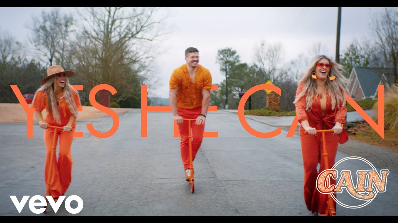
Watch the official music video for “Yes He Can” now!
Listen to #YesHeCan now: https://CAIN.lnk.to/RiseUpAlbumID!CFVIZ
Tour Dates - https://CAIN.lnk.to/TourDatesID
Church Resources - https://CAIN.lnk.to/ChurchResourcesID
Connect with us:
Email list: https://CAIN.lnk.to/EmailListID!RiseUpMV!YHCMV
Facebook: https://CAIN.lnk.to/FacebookID!YHCMV
Instagram: https://CAIN.lnk.to/InstagramID!YHCMV
Twitter: https://CAIN.lnk.to/TwitterID!RiseUpMV!YHCMV
Website: https://CAIN.lnk.to/OfficialWe....bsiteID!RiseUpMV!YHC
Yes He Can
Sometimes I wonder, is He faithful?
Does He see me in my trouble
Does He understand
Sometimes I question if He’s able
Can He rescue, can He save me
Again and again
But when I look back
Did He move every mountain?
Did He part every sea?
Yes He did
So yes He can
Did He defeat the darkness?
Did He deliver me?
Yes He did
So yes He can
Yes He did
So yes He can
Sometimes those voices try to tell me
I’m forgotten, and I’ve fallen
Too far from His hands
But I know what kind of God He is
And I’m trusting in His promises
I’m believing
And I’m singing
Yes He can
Cause I’ve seen too much
Now I can’t deny
He’s come through
Every single time
From the beginning until the end
He did, He will, He can
Music by CAIN performing “Yes He Can” (C) 2021 Provident Label Group LLC, a division of Sony Music Entertainment
#CAIN #ChristianMusic #YesHeCan

Beautiful worship for children! The perfect music to play for your children to see them praise God!
Tracklist:
What A Beautiful Name
Little Life
Oceans (Where Feet May Fail)
Who You Say I Am
You Know Me
You Say
Be Still
Tiny Little Voice
To Be Like You
This I Believe
Thank You Jesus
O Praise The Name
Nothing LIke Your Love
Jesus You Care
Sinking Deep
Gave It All
#kidmin #kidsworship #worship
Listen to our new album:
https://hillsong.lnk.to/canyoubelieveit
*******Follow Us*******
hillsongkids.com
hillsongkidsbig.com
TWITTER - http://twitter.com/hillsongkids
FACEBOOK - https://facebook.com/hillsongkids
INSTAGRAM - http://instagram.com/hillsongkids
-------------------------------------
Engaging children of this generation in worship - - Building the lives of children all over the globe.
We believe that teaching children to love God and others takes place in both the home and in the church. For this reason we seek to partner with parents (the greatest teachers of all!) and with church pastors and leaders, equipping them with great resource. Through fun experiences, meaningful music and ministry that encourages kids to participate, we present Jesus Christ and His Church in a relevant way, creating moments which children will never forget.
Anyone can do what we do. The simple key is to love God, love His Church and have fun!

---------------------------------------------------
🔻Follow "Praise Worship Music"
➞ Top Vidoes Hillsongs: https://bit.ly/3xbFxir
➞ Subscribe for More: https://bitly.com.vn/j6ar4y
✚Please share this video in social sites (Facebook, Google +, Twitter.)✚
📌 Facebook: https://bitly.com.vn/zvzxcl
📌 Soundcloud: https://bitly.com.vn/qgmtfj
📌 TikTok: https://bitly.com.vn/pthcga
📌 Instagram: https://bitly.com.vn/g75awh
We hope to hear your comments
#worshipsongs #christiangospel #praiseandworship
---------------------------------------------------
► Music and Video Copyright belongs to @Praise Worship Music. DO NOT reupload, otherwise, you will get copyright strikes.
Sincere thanks: Praise Worship Music Team !
---------------------------------------------------
►Tag:
worship,praise,praise and worship,,gospel music,gospel topic,worship songs,praise worship songs,worship and praise songs,worship songs 2021,gospel songs ,christian songs and praise,praise songs,christian songs 2022 ,latest gospel music,christian songs with lyrics,christian music,best worship songs gospel songs,christian songs ,worship songs with lyrics,praise and worship songs with lyrics
#praiseworshipsongs #praiseandworship #worship
#worshipsongs #BestPraiseWorship #praiseandworshipsongswithlyrics
#Praise
#Christian
#Gospel
#worshipsongswithlyrics
#worshipsongsofhillsong
#worshipsongslyrics
#worshipsongsever
#worshipsongscollection
#worshipsongsalltime
#worshipsongs
#worshipmusicchristian
#worshipmusic2021
#worshipmusic
#worshipgospelmusic
#worshipandpraisesongs
#worship2021
#worship
#wepraiseyou
#topworshipsongs
#toppraiseandworshipsongs
#top100praiseandworshipsongs2021
#top100christianworshipsongs
#top100bestchristiangospel
#prayers
#prayersongslyrics
#prayersongs2021
#prayersongs
#praiseworshipsongs
#praiseworshipmusic
#praisethelord
#Lordineedyou
#praisesongs
#praiselord
#praiseandworshipsongs2021
#praiseandworshipsongs
#praiseandworshipmusic
#praiseandworship
#praiseandhymns
#praise&worshipsongs2021
#praise
#powerfulpraiseandworshipsongs
#nonstopworshipsongs
#nonstopchristiansongs
#nigeriangospelmusic
#nigeriangospelmix
#newworshipsongs
#newpraiseandworshipsongs
#newchristiansongs
#newchristian
#musicpraise
#morningworshipsongs2021
#morningworshipsongs
#morningworship
#morningpraiseandworshipsongs
#morningworshipsongs
#louvoresdeadoração
#laurendaigleworshipsongs
#laurendaiglesongs
#laurendaigleplaylist
#laurendaiglefullalbum
#laurendaiglealbums
#laurendaigle
#latestpraisesongs
#latestgospelsongs2021
#latestgospelsongs
#latestgospelmusic
#latestchristiansongs2021
#latestchristiangospel
#hymnssongspraise
#hymnssongs
#hymnsoffaith
#hymns
#hillsongspraiseandworshipsongsplaylist
#hillsongworshiptoptracks
#hillsongworshipsongs
#hillsongworshiplyrics
#hillsongworship2021
#hillsongworship
#hillsongunited
#hillsongtoptracks
#hillsongsongslyrics
#hillsongsongs
#hillsongpraiseandworshipsongs
#hillsongplaylist
#hillsongmúsica
#hillsongmusic2021
#hillsongmusic
#hillsonghits
#hillsongfullalbum
#hillsong2021
#hillsong
#highworshipsongs
#greatestworshipmusic
#gospelworshipsongs
#gospeltopic
#gospelsongswithlyrics
#gospelsongs2021
#gospelsongs
#gospelmusicpraiseandworshipsongs
#gospelmusicpraiseandworship
#gospelmusicafrica
#gospelmusic2021
#gospelmusic
#gospelcountrymusic
#gospelchristiansongs
#gospel2021
#gospel
#goodpraisesongs
#globalgospelgroup
#dunamischurch
#countrygosspel
#countrygospelsongs
#christianworshipsongs2021
#christianworshipsongs
#christiansongswithlyrics
#christiansongsnonstop
#christiansongslyrics
#christiansongsandpraise
#christiansongs2021
#christiansongs
#christianmusic 2021
#christianmusic
#christianhyms
#christiangospel
#christian
#bestworshipsongsever
#bestworshipsongs
#bestworshipmusic
#bestpraiseworship
#bestpopularworshipsongs2021
#besthymns
#bestchristiansongs2021
#200praiseandworshipsongs
#100praiseandworshipsongs
#hillsong
#churchonline
#hillsongchurch
#hillsongmusic
#sundayworshiponline
#asitisinheavenhillsongchurch
#soletitbemyheartcriesholy
#theearthcriesholy
#2021worshipnosicknessinmybody
#noprisonwallscanholdme
#thefearlesslightofglory
Short Good Morning Prayers To Use On A Daily Basis


Spirit Filled and Soul Touching Gospel Worship Songs for Prayers | ►Download Link: https://josephkokumu.com/ts22 | DJ Lifa #TotalSurrender22 |
►Subscribe https://bit.ly/LifaYouTube
►Love this mix? Get similar mixes through this link: https://bit.ly/englishMixes
**************
More Playlists
►All DJ Lifa Mixes: https://bit.ly/DJLifaMixes
►English Worship Mixes https://bit.ly/SwahiliMixes
►Reggae Praise and Worship Mixes: https://bit.ly/DJLifaReggae
►Congolese Praise and Worship Mixes: https://bit.ly/CongoleseMixes
►Instrumentals for Worship | Prayer | Relaxation | Rest ( Produced by DJ Lifa ): https://bit.ly/DJLifaBeats
►Video Mixes: https://bit.ly/VideoMixes
►Music Written by Joseph Kokumu (DJ Lifa): https://bit.ly/DJLifaSongs
**********************************************
► Get Nothing is Impossible by @Faith Lumumba (Written by DJ Lifa ( Joseph Kokumu) ) https://lifarecords.co.ke/nifl
**********************************************
***********************************************
Follow DJ Lifa
►Facebook: https://facebook.com/djlifamusic
►Instagram: http://instagram.com/djlifamusic
►Twitter: https://twitter.com/djlifamusic
***************************
For mixes, visit any of the sites below
►Mixcloud: https://mixcloud.com/deejaylifa
►Vimeo: https://vimeo.com/deejaylifa
►Mdundo: https://mdundo.com/a/17866
►YouTube: https://youtube.com/c/djlifa
►DJ Lifa Website: https://josephkokumu.com/dj-mixes
************************************************
For Media Interviews, Partnerships, Commercials, Appointments, Events & Bookings, Please Call 254769757859 or Email: info@josephkokumu.com /deejaylifa@gmail.com
►Website: https://josephkokumu.com
************************************************
Tracklist
0:00 GUC - All That Matters https://www.youtube.com/watch?v=-r6mL7Ws0Cc
05:52 MOG Music - Elohim https://www.youtube.com/watch?v=7eAvIYagrrs
11:49 Toby - Amen https://www.youtube.com/watch?v=dcMaP2mzDGw
15:53 MOG Music - Be Lifted https://www.youtube.com/watch?v=yH1FJEQBzss
23:45 Judikay - Yes Lord (LIVE) https://www.youtube.com/watch?v=KZ3YJ2wNYQM
30:15 Moses Bliss - Too Faithful https://www.youtube.com/watch?v=YA2Lxfw4SSw
35:23 Joe Mettle - Wonderful, Merciful Saviour https://www.youtube.com/watch?v=dCXywtu_vIc
40:44 Evelyn Wanjiru and Eunice Njeri - Worthy https://www.youtube.com/watch?v=pVflrLid7T0
44:00 Evelyn Amo - Lift His Name Higher https://www.youtube.com/watch?v=qNuPtFFPoLs
47:47 Joe Mettle - Come Holy Spirit https://www.youtube.com/watch?v=rgGIg0BEsiU
52:39 MOGMusic ft. Ps Edwin Dadson - God of Miracles https://www.youtube.com/watch?v=K69jtKXWcvU
57:02 Collen Maluleke - You are Glorious https://www.youtube.com/watch?v=dde-Gw-w8Bk
1:02:44 GUC - I'm Yours https://www.youtube.com/watch?v=Ibtd6FVDrXs
1:08:46 Onos - Glorify Emmanuel https://www.youtube.com/watch?v=Wt9PsKjQo8E
1:12:31 Chris Shalom - Bigger https://www.youtube.com/watch?v=zXM2bVPbCJ4
1:16:12 Denzel Prempeh - Yes You are the Lord https://www.youtube.com/watch?v=MpCKY9WfaA8
1:22:00 Isabel Davis - Wide as the Sky https://www.youtube.com/watch?v=5i4QL9ZpFJI
1:29:02 Sound of Heaven Worship - Draw me Close to You & Jesus at the Center https://www.youtube.com/watch?v=Fl7899v18H4
1:39:25 Hillsong Worship - Glorious Ruins and Anchor Mash Up https://www.youtube.com/watch?v=s-ZGNYTu6Vk
1:45:41 Judikay - Holy Ghost https://www.youtube.com/watch?v=NkVqejjnQ2A
1:52:49 Prospa Ochimana - You are worthy to be Praised https://www.youtube.com/watch?v=lw0qvQGBbuk
1:57:28 JudiKay - More than Gold https://www.youtube.com/watch?v=AEGjZ2ERKnU
This mix is provided for promotional purposes only.
____________________________________________
#TotalSurrender
Enjoy this African Worship mix by DJ Lifa featuring some of the best African English Gospel Music Praise and Worship.
Subscribe and check out more DJ Lifa mixes for the best spirit-filled worship songs, Christian songs, Christian songs 2021, Christian music 2021, gospel songs, best worship songs, Christian music, worship music, worship songs 2021, best worship music, praise and worship, praise and worship music, praise worship songs, praise,worship, worship songs, gospel music 2021, latest Christian gospel, morning worship songs, morning prayers, prayer song, gospel songs 2021, new worship, new Christian, greatest Christian
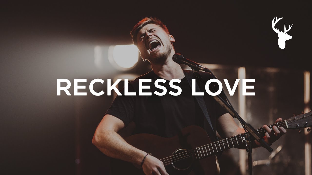
Cory Asbury singing "Reckless Love" at Heaven Come Conference 2017
Subscribe to our channel for weekly videos: http://bit.ly/BMsubscribe
Buy or Stream the official single here: https://BethelMusic.lnk.to/RecklessLoveID
Chord Chart: https://bethelmusic.com/chords....-and-lyrics/reckless
CCLI: https://songselect.ccli.com/So....ngs/7089641/reckless
Connect with Cory:
https://www.facebook.com/coryasburymusic/
https://www.instagram.com/coryasbury/
https://twitter.com/REALCORYASBURY
https://bethelmusic.com/artists/cory-asbury/
Lyrics:
Reckless Love
Written by Cory Asbury, Caleb Culver, and Ran Jackson
Verse 1
Before I spoke a word, You were singing over me
You have been so, so good to me
Before I took a breath, You breathed Your life in me
You have been so, so kind to me
Chorus
Oh, the overwhelming, never-ending, reckless love of God
Oh, it chases me down, fights ‘til I’m found, leaves the ninety-nine
I couldn’t earn it, I don’t deserve it, still You give Yourself away
Oh, the overwhelming, never-ending, reckless love of God
Verse 2
When I was Your foe, still Your love fought for me
You have been so, so good to me
When I felt no worth, You paid it all for me
You have been so, so kind to me
Bridge
There’s no shadow You won’t light up
Mountain You won’t climb up
Coming after me
There’s no wall You won’t kick down
Lie You won’t tear down
Coming after me
